Degenerative Joint Disease (DJD)
DJD is a progressive degeneration of articular cartilage in a synovial joint occurring in 3 stages. synovitis, arthritis and finally osteoarthritis. Affecting the capsular ligament both outer and inner layers, the synovial fluid and articular cartilage.
The first stage of synovitis is an inflammation synovial structures causing heat swelling and pain. The articular cartilage becomes damaged by this DJD and has a loss of function. The synovial structures release enzymes causing more damage, this creates a vicious cycle. This inflammation stretches the capsular ligament which again increases synovial production (more swelling)> When these enzymes are released they are less viscous than the synovial fluid meaning that this mix is less lubricating and the function is lost somewhat.
The arthritis is the inflammation of the whole joint due to the synovitis. As the synovial fluid becomes thinner than the articular cartilage is no longer nourished or lubricated as it was, it is slowly worn away with excessive work, thinning the cartilage down making it less resistant and shock absorbing making the problem worse. Lysosomal enzymes are then released from the breaking down cartilage causing a further reduction in use of the synovial fluid leading to a further breakdown of the cartilage its self. Now because there is no direct blood flow to this cartilage and it gets it from the synovial fluid the cartilage cannot repair, this failure to repair leads to major cartilage damage and leading to bony changes, this is called osteoarthritis.
Osteoarthritis is the final stage of DJD and involves everything talked about plus the sub chondral bone. This means bone remodelling due to excessive compression. exostosis of joints, narrowing of joint space, inflammation of bone causing fractures. This is going to create severe pain for the horse, commonly seen in stiffness and lack of want to do any work. This in turn can lead to lack of muscle, this makes the problem worse…. It is all one nasty cycle that continues to loop.
Falling, interfering, getting kicked can all be traumatic injuries that directly relate to this process starting. Poor conformation again another very common predisposing factor. over worked on hard grounds and general wear and tear will all come to aid this process. Often seen in the aging horse often seen as just a part of growing older. Farriery can sometimes be to blame, leaving feet unbalanced can cause that uneven loading the same as poor conformation which will lead to swelling or synovial structures and if left unattended the DJD kicks off.
As discussed signs often show in swelling and lameness, abnormal shoe wear can be seen, loss of function and changes in synovial fluid (veterinary diagnoses). Often the diagnosis is through observation and x-rays often to follow will make the conformation of your thoughts correct.
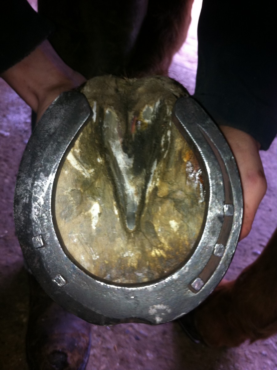
Making the horse comfy is the only treatment, rest for the horse, anti-inflammatory, corticosteroids or hyalauric acid injections to joint are the only options, there is arthrodesis although uncommon and not widely practiced, and fusing the joints in this process eliminates the painful process. For farriers creating even loading is essential, creating level foot fall for joints and returning HPA to normal. This can all be done with aid of lateral and DP x-rays to help you assess what to trim. Reducing break over of shoe is often promising, knocking the edge of all the way around is simple yet very affective, and this allows the foot to break over where it is comfortable.
Unfortunately as a progressive disease prognosis is usually very poor.
Ringbone (high and low Articular and Peri-Articular)
Ringbone is a process of new bone growth formation involving all the phalanxes, it is described by its position and whether the joint is involved. High ring bone involves distal P1 and proximal P2 and low ring bone is distal P2 and proximal P3. Articular ringbone involves the joint hence the articulation and the peri-articular is around the joint capsule but doesn’t affect the joint. You can have both articular and peri-articular ringbone in the one horse. Often more seen in the fronts due to the excessive concussion and 60% weight bearing.
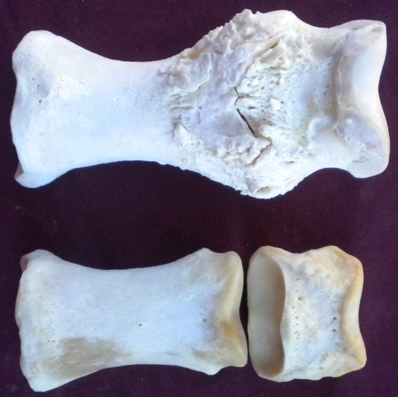 Ringbone is exostosis due to tension on the periosteum where ligaments, a joint capsule or tendon attach. Tearing of extensor tendons on insertion points causes the damage and whilst healing the tendinous structures turn to bone causing these osseous ridges seen.
Severe low ringbone can produce a distinct hard bony swelling at the coronary band sometimes referred to as pyramidal disease. Can be seen as intermittent lameness with heat and swelling in the pastern areas depending on the ringbone being high or low. Feeling bone growth can be quite tricky though as soft tissues covering the bone make it almost impossible to feel however palpation of the areas may produce a pain reflex.
Sometimes true and false ringbone can be used, true ringbone is new bone growth across a joint which is articular because it limits articulation. Proximal and middle phalanx bone growth not involving the joint is false ringbone. False ringbone seen more in cobs and heavy horses with upright feet as its more concussion based and the false ringbone is seen in fast turning horses like polo or show jumping.
To properly diagnose this must be x-rayed to see the bony changes, bearing in mind that in early stages the bone growth may be hard to see as its much lighter than the bone already there. MRI or scans can be used that show up the bony changes better. As you’ll see from the x-rays the ridges on these bones can cause severe irritation to soft tissue structures. On the front the extensor tendons may be affected especially if the bone growth is near the attachment points. On the palmar/plantar surfaces the suspensory ligament is at risk from irritation.
The vet and farrier aim to reduce movement in the joint as that causes the pain, vets are likely to administer anti-inflammatory pain killers and offer shoeing advice. A omnidirectional break over shoe allows the horse to get its foot away where it is comfortable which is what we most commonly see also the uses of rocker bar shoes are used to make the rocking motion another joint taking the articulation away from the affected ringbone joint.
Prognosis is fair but return to work will be a lower standard, there will be zero movement in this joint once all fused but there will be no pain after joint has fused.
Sidebone (uni-lateral & bi-lateral)
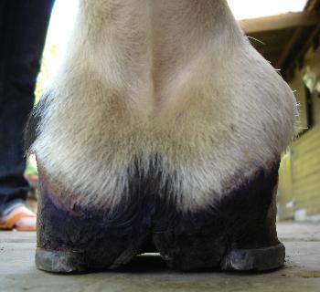
Side bone is ossification of the lateral cartilages characterized by loss of full biomechanical function of the equine. Forelimbs more commonly seen as its more weight bearing and this disease is a concussion based ailment. Broken down unilateral side bone affects one side of the foot (one of the lateral cartilages) and bi lateral side bone is affecting both lateral cartilages in the hoof. Remember that effectively you have a medial and a lateral, lateral cartilage, sometimes referred to as the collateral cartilages.
 Causes are usually down to conformation as this makes uneven loading causing excess stress on joints leading to ossification trying to harden up the area being hit harder. Type of work done and road work will produce more concussion, also uneven loading which can be caused by roadwork as the have uneven cambers. Direct trauma can also cause sudden ossification of the cartilages. On top of this overweight horses, over weight owners are all helping to increase the load. Poor management leaving the feet to long to be shod so they go unbalanced again leading to uneven load. Lastly poor farriery can also cause unlevel footfall by poor trimming which causes more uneven loading.
Typical signs of this will be uneven shoe wear, unilateral side bone can be seen as massive wear to the outside branch and bilateral side bone will destroy the toe very quickly to wear it is comfortable, seen as pawing in the stables to scrape it away until the horse gets comfortable. There may be a change in gait and foot fall becoming unlevel. There will be mild lameness present when ossification takes place and there will be a lack of elasticity of the cartilage leading to a lack of expansion and function of the foot. Bulging in the coronary band with coronary shunting may become visible because of the foot becoming more upright due to ossification which is restricting expansion. Heat and swelling may also be obvious.
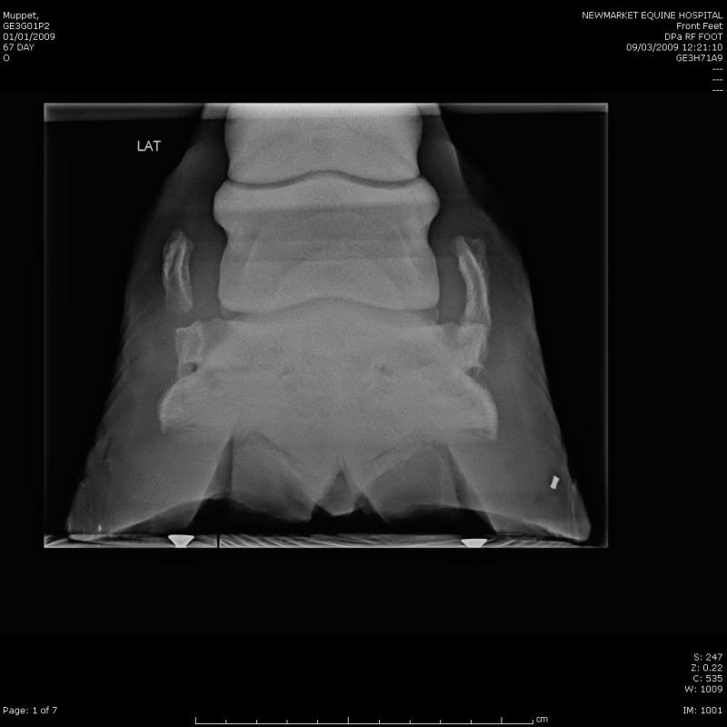 Other diagnoses will be need to done as an x-ray to fully see the extent of side bone. The x-rays needed are usually a lateral medial or sometime a dorso palmar.
When ossified there shouldn’t be a problem unless the now bony cartilage fractures and snaps of causing an infection. This is known as a quitter and will have to be surgically removed. Depending on where this is will depend on how easy or hard removing of this fragment will be.
 Use of anti-inflammatory drugs to ease pain but turn out will be the best option and allow it to run its course. If horses are still in work then a side bone shoe can be used by taking of the branch and making 2 nail holes right near the toe to promote expansion of the upright quarter and allow easy break over reducing the pain. In bilateral cases a graduated shoe is used. But again the best remedy is turning the horse out for 6 months and allowing the process to finish. Achieve level footfall with your trim to create even loading
Prognosis is very good when the ossification stops as pain goes away. Although the loss of function to the foot will now always be present.
Bone Spavin
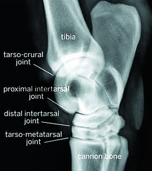
Bone spavin is osteoarthritis or the final phase of DJD in the lower three hock joints. Usually affecting the lowest two joints called the tarsometatarsal and distal inter tarsal joints. Seen in older mature horses although it’s not unseen in youngsters from time to time. Seen in jumping horses or ones that turn sharply. Poor conformation leading to this is usually cow hocked or sickle hocked.
 Cartilage compression overtime between upper and lower surfaces of the lower tarsal bones to become compressed and eroded, the joint spaces become smaller and new bone growth will occur.
Uneven loading causes excessive compression of the cartilage and bone on one side and strain in the joint capsule and supporting ligaments on the other side. Repeated overloading on one side causes exostosis to occur. The other contributing factors are as discussed earlier such as cow or sickle hocks. Dressage, jumping, hunting and racing all cause excessive hock flexion and repeated loading strain. Juvenile spavin is the occurrence of bone spavin in horses less than 3 years old occurring before the horse as done much work. Osteochondritis lesions are likely cause in some cases it can also be a secondary to distortion of cuboidal bones in premature foals.
Diagnoses can be seen in x-rays and the scraping of the toes on the hind feet as the hock fails to flex properly.
Drugs will be administered by a vet to reduce pain and swelling. Corticosteroid or nerve blocks can be used but have to be careful to comply with competition rules as this will not be permitted in most. Shoeing is very important and must help break over with rolled toes and wedges if necessary. Shoe with heel supports such as egg bars are very good as the support the hock with their width and length. Even foot fall must be achieved for stress on the hock to be eased.
Advice to the owners of exercising daily is essential preferably ridden work as lunging places uneven stress due to not being a straight line. Surgery may be considered as arthrodesis moves the fusion along ending the lameness.
Guarded prognosis as it depends on severity and joints involved, use of the horse and how quickly its deteriorating. Competing at high level won’t last long but pleasure riding should be achievable.